Brain MRI
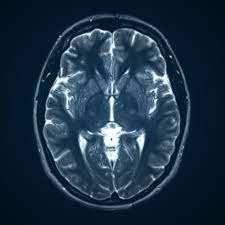
What does an MRI of the brain show?
A brain MRI can help doctors look for conditions such as bleeding, swelling, problems with the way the brain developed, tumors, infections, inflammation, damage from an injury or a stroke, or problems with the blood vessels. The MRI also can help doctors look for causes of headaches or seizures
How long does an MRI of the brain take?
A brain MRI can take about 30 minutes to an hour to complete. It may take longer if you’re getting a brain MRI with contrast. Your healthcare provider will be able to give you a more exact time range based on the specific reason for your scan
Why would a neurologist order a brain MRI?
MRI is used to diagnose stroke, traumatic brain injury, brain and spinal cord tumors, inflammation, infection, vascular irregularities, brain damage associated with epilepsy, abnormally developed brain regions, and some neurodegenerative disorders
How do you prepare for a brain MRI?
Before an MRI exam, eat normally and continue to take your usual medications, unless otherwise instructed. You will typically be asked to change into a gown and to remove things that might affect the magnetic imaging, such as: Jewelry. Hairpins
Is MRI better than CT scan for brain?
Advantages of MRIs
Magnetic resonance imaging produces clearer images compared to a CT scan. In instances when doctors need a view of soft tissues, an MRI is a better option than x-rays or CTs. MRIs can create better pictures of organs and soft tissues, such as torn ligaments and herniated discs, compared to CT images
Magnetic resonance imaging produces clearer images compared to a CT scan. In instances when doctors need a view of soft tissues, an MRI is a better option than x-rays or CTs. MRIs can create better pictures of organs and soft tissues, such as torn ligaments and herniated discs, compared to CT images
Does brain MRI have side effects?
On very rare occasions, a few patients experience side effects from the contrast material. These may include nausea, headache, and pain at the site of injection. It is very rare that patients experience hives, itchy eyes, or other allergic reactions to the contrast material
What not to do before an MRI of the brain?
You can prepare for your scan ahead of time by removing the following items from your body and pockets:
- Body piercings.
- Pens.
- Jewelry.
- Hearing aids.
- Pins.
- Hairpins.
- Zippers or any metal clothing fasteners.
- Removable dental work.
Can brain MRI show nerve damage?
Does an MRI scan show nerve damage? A neurological examination can diagnose nerve damage, but an MRI scan can pinpoint it. It’s crucial to get tested if symptoms worsen to avoid any permanent nerve damage

Does brain MRI need empty stomach?
On the day of your MRI scan, you should be able to eat, drink and take any medication as usual, unless you’re advised otherwise. In some cases, you may be asked not to eat or drink anything for up to 4 hours before the scan, and sometimes you may be asked to drink a fairly large amount of water beforehand.
Why do I feel dizzy after MRI?
In a new study published in Current Biology online on Sept. 22, a team led by Johns Hopkins scientists suggests that MRI’s strong magnet pushes on fluid that circulates in the inner ear’s balance center, leading to a feeling of unexpected or unsteady movementc
Can MRI show blood clots?
Magnetic resonance imaging (MRI) may be used to diagnose deep vein thrombosis (DVT) in patients for whom ultrasound examination is inappropriate or unfeasible. We undertook a systematic review of the literature and meta-analysis to estimate the diagnostic accuracy of MRI for DVT.
Why can’t I drink water before an MRI?
If You Have an Overactive Bladder
This feeling of urgency can make it harder to hold urine in. While you may still experience this urgency to a degree, not drinking for several hours before your procedure can make you less likely to experience incontinence during the scan.
This feeling of urgency can make it harder to hold urine in. While you may still experience this urgency to a degree, not drinking for several hours before your procedure can make you less likely to experience incontinence during the scan.
Why do they inject dye for an MRI?
During the procedure, they’ll inject the gadolinium-based dye into your arm intravenously. The contrast medium enhances the image quality and allows the radiologist more accuracy and confidence in their diagnosis.
Can brain MRI cause vertigo?
Vertigo induced by exposure to the magnetic field of a magnetic resonance imaging (MRI) scanner is a well-known phenomenon within the radiology community but is not widely appreciated by other clinical specialists
Is MRI dye painful?
Most people feel nothing after receiving an injection of contrast, Dr. Taouli says. Only a very small number of people will have adverse effects. Of those, a skin rash, hives, and pain at the injection site are more common.
Can contrast dye affect your brain?
We hypothesised that the direct neurotoxic effect of the contrast agent also contributed to the patient’s progressive and fatal brain oedema. All types of iodinated contrast agents can induce the development of neurotoxicity, but the occurrence of fatal cerebral oedema is very rare.
Is MRI contrast harmful to kidneys?
The type of gadolinium used in older contrast agents isn’t safe for people with moderate or advanced chronic kidney disease. Older versions of contrast agents that contain gadolinium increase the risk of a rare but serious disease called nephrogenic systemic fibrosis.