Understanding the Lumbar Spine: A Guide to MRI Scans

Introduction to the Lumbar Spine
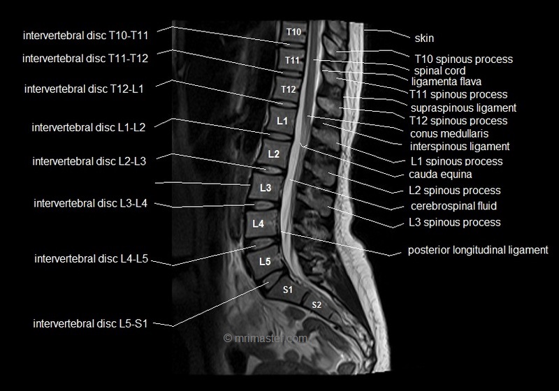
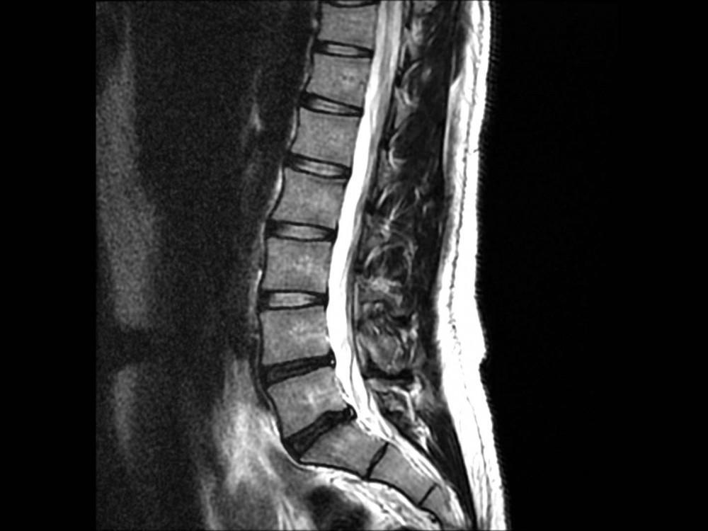
The lumbar spine, also known as the lower back, is a complex structure of bones, muscles, ligaments, and nerves that support the weight of the upper body and allow us to stand upright, bend, twist, and move. It consists of five vertebrae (L1-L5) that are stacked on top of each other and separated by intervertebral discs that act as shock absorbers.
How MRI Scans Work?
During an MRI scan of the lumbar spine, the patient lies on a table that slides into a tunnel-shaped machine. The machine generates a strong magnetic field that causes the hydrogen atoms in the body’s tissues to align in a certain way. Radio waves are then used to disturb the alignment of these atoms, which emit signals that are picked up by the machine and processed into images.
Benefits of MRI Scans
One of the main advantages of MRI scans is that they are non-invasive and do not use ionizing radiation, which can be harmful to the body. MRI scans can also produce highly detailed images of the lumbar spine, allowing doctors to detect abnormalities that may not be visible on X-rays or other imaging tests.
Risks and Precautions
Although MRI scans are generally safe and well-tolerated, there are some risks and precautions that patients should be aware of. For example, patients with metal implants or pacemakers may not be able to undergo an MRI scan, as the magnetic field can interfere with these devices. Patients who are claustrophobic or anxious may also experience discomfort or anxiety during the scan.
Conclusion
MRI scans are a valuable tool for diagnosing and evaluating conditions that affect the lumbar spine. By providing detailed images of the internal structures of the body, MRI scans can help doctors make accurate diagnoses and develop effective treatment plans. If you are experiencing back pain or other symptoms of lumbar spine problems, talk to your doctor about whether an MRI scan may be right for you.
Q&A
Q: What is a lumbar spine MRI? A: A lumbar spine MRI is a non-invasive imaging test that uses a powerful magnetic field and radio waves to produce detailed images of the bones, discs, muscles, and nerves in the lower back.
Q: Why is a lumbar spine MRI performed? A: A lumbar spine MRI is performed to help diagnose and evaluate conditions that affect the lower back, such as herniated discs, spinal stenosis, degenerative disc disease, and tumors.
Q: What happens during a lumbar spine MRI? A: During a lumbar spine MRI, the patient lies on a table that slides into a tunnel-shaped machine. The machine generates a strong magnetic field that causes the hydrogen atoms in the body’s tissues to align in a certain way. Radio waves are then used to disturb the alignment of these atoms, which emit signals that are picked up by the machine and processed into images.
Q: Is a lumbar spine MRI painful? A: No, a lumbar spine MRI is not painful. However, some patients may experience discomfort or anxiety during the scan, especially if they are claustrophobic or have difficulty lying still for an extended period of time.
Q: How long does a lumbar spine MRI take? A: A lumbar spine MRI typically takes 30-60 minutes to complete, depending on the type of scan and the number of images that need to be taken.
Q: Are there any risks associated with a lumbar spine MRI? A: Although lumbar spine MRI is generally considered safe, there are some risks and precautions that patients should be aware of. For example, patients with metal implants or pacemakers may not be able to undergo an MRI scan, as the magnetic field can interfere with these devices. Additionally, there is a small risk of an allergic reaction to the contrast dye that is sometimes used during the scan.
Q: What should I expect after a lumbar spine MRI? A: After a lumbar spine MRI, you can resume your normal activities immediately. Your doctor will review the images and discuss the results with you at a follow-up appointment. Depending on the findings, further tests or treatment may be recommended.
Q: How can I prepare for a lumbar spine MRI? A: To prepare for a lumbar spine MRI, you should wear loose, comfortable clothing and avoid wearing jewelry or metal objects. You may also be asked to fast for a few hours before the scan, especially if contrast dye will be used. It’s important to inform your doctor if you have any metal implants or devices, as these may need to be removed before the scan.